Which X Ray Would Be Used to Visualize the Gallbladder
A series of X-rays are taken of the gallbladder after you swallow a special contrast dye. The test was once the standard for diagnosing diseases of the GB such as gallstones but is used less frequently now.

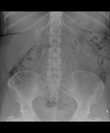
Abdominal X Ray Showing Two Plastic Stents Double Pigtail In The Gall Download Scientific Diagram
What X-ray would be used to visualize the gallbladder.
. Which X-ray would be used to visualize the gallbladder. Which x-ray would be used to visualize the gallbladder. A physician may order an X-ray to check for certain cancers in different parts of the body by.
The study involves taking tablets containing dye contrast which outline any abnormalities when x rays are taken the following day. The x-ray technologist should be informed since the gallbladder might not be well visualized. Results A normal OCG will show a normal gallbladder.
The function of the gallbladder is to store. Oral cholecystogram is an X-ray imaging procedure used to examine the gallbladder a sac-like organ in the right upper abdomen that stores bile before it is released through the bile ducts into the small intestines to help digest fat. This is a test that uses X-rays and a computer to make detailed images of the body.
Bite-wing- X-ray X-ray taken with a part of the film holder held between the teeth and parallel to the teeth. See answer 1 Best Answer. Which X ray would be used to visualize the gallbladder A IV cholecystography B.
The gallbladder should visualize and be free of any solid structures such as stones polyps or tumors. Which X-ray would be used to visualize the gallbladder. School New York University.
Course Title RELIGIOUSS 2597. Some X-ray types require that a patient swallow a dye or have dye injected into the body so the X-ray can capture a. It should empty freely with no obstruction after the PFM post-fatty meal.
A gallbladder scan is a type of nuclear. A gallbladder scan is a specialized radiology procedure used to assess the function and structure of the gallbladder. An upper GI series looks at the esophagus stomach and duodenum.
X-rays are used for a multitude of reasons. What type of X-ray would be used to visualize the gallbladder. A cholangiography examines the hepatic duct common bile duct and pancreatic duct.
Which of the following endoscopic exams would be used to view inside a section of the colon. Which x ray would be used to visualize the gallbladder. This condition is somewhat uncommon and results from calcification of the gallbladder wall.
Rarely in gravely ill patients x-rays show air in the biliary tree which suggests emphysematous cholangitis. This is an x-ray exam of the gallbladder GB a sac-like organ that stores bile that is located under the liver. A lower GI series examines the colon and rectum.
This test can show gallstones inflammation of the gallbladder cholecystitis and other problems. You could need other tests along with an x-ray. Upper GI series c.
A simple abdominal x-ray can be used to identify calcified gallstones. What type of X-ray would be used to visualize the gallbladder. This procedure may also be referred to as a liver-biliary scan because the liver often is examined as well due to its proximity and close functional relationship to the gallbladder.
How the test is performed. Which abbreviation stands for a pathological condition. A gallbladder ultrasound is a noninvasive and typically painless examination used to diagnose conditions related to the gallbladder.
Your health provider may order an x-ray if they suspect gas buildup calcified gallstones or porcelain gallbladder concerns. Reasons for a Gallbladder X-Ray. Cholecystogram X-ray image of the gallbladder.
Which x ray would be used to visualize the. Which medication is used to treat constipations. A lower GI series examines the colon and rectum.
An IV cholecystography is an X-ray of the gallbladder after intravenous dye is administered. An intravenous cholecystography is an X-ray of the gallbladder after intravenous dye is administered. An upper GI series looks at the esophagus stomach and duodenum.
In some cases you may need to swallow a substance called iopanoic acid or receive it as an injection. Peptic ulcer disease occurs in the. Sometimes patients are then asked to drink a highfat formula that will cause the gallbladder to contract and release bile.
Which X-ray would be used to. A cholangiography examines the hepatic duct common bile duct and pancreatic duct. Because only 10 of all gallstones are calcified this imaging study has limited usefulness.
Intravenous cholecystography Dye is administered intravenously to the patient allowing for X-ray visualization of the gallbladder and bile ducts. An X-ray of the gallbladder can be viewed through an abdominal X-ray which sometimes requires the patient to swallow a dye or have it injected into the body in order to create a clearer image of the gallbladder. An abdominal X-ray can spot gas and some types of gallstones containing calcium.
Porcelain gallbladders can also be seen in plain x-rays. Pages 79 This preview shows page 24 -. MRI scan is used to scan gallbladder.
The following day at the hospital the radiologist examines the gallbladder with a fluoroscope a special x ray that projects the image onto a video monitor. Lower gastrointestinal series lower GI series. Plain x-rays can detect a calcified porcelain gallbladder.
Magnetic resonance imaging MRI MRI is used to image blood vessels without using. But this test isnt used often. The procedure for getting an x-ray can vary.

Which Test Is Best For The Detection Of Gallstones An Ultrasound An Mri Scan Or A Ct Scan Quora
Gallstones Radiology Reference Article Radiopaedia Org
2
No comments for "Which X Ray Would Be Used to Visualize the Gallbladder"
Post a Comment